

 0
0
 0
0
■ 1. Background
LCL161 (T2080), CAS: 1005342-46-0 is a small-molecule SMAC mimetic that functions as a pan-IAP antagonist with oral activity, designed to disengage inhibitor of apoptosis proteins (IAPs) from their control over caspases and TNF receptor signaling. In cellular assays, LCL161 inhibits XIAP with an IC50 of 35 nM in HEK293 cells and potently inhibits cIAP1 with an IC50 of 0.4 nM in MDA-MB-231 cells, demonstrating subfamily-selective potency that is well aligned with the canonical SMAC–IAP interaction interface. By mimicking the N-terminal AVPI motif of endogenous mitochondrial SMAC/DIABLO, LCL161 binds the BIR domains of IAPs, thereby displacing effector and initiator caspases from XIAP and relieving direct proteolytic inhibition of caspase-3, -7, and -9 [1].
Within the IAP signaling axis, XIAP acts primarily as a direct caspase inhibitor, whereas cIAP1 and cIAP2 serve as E3 ubiquitin ligases that orchestrate receptor-proximal complexes downstream of TNF receptor 1 (TNFR1), controlling RIPK1 ubiquitination, NF-κB activation, and cell survival decisions [2]. Consistent with this pathway architecture, LCL161 antagonism of cIAP1 promotes its rapid auto-ubiquitination and proteasome-dependent loss, shifting TNFR1 signaling away from ubiquitin-dependent pro-survival Complex I toward cytosolic death-inducing platforms that favor caspase-8 activation. Concomitant XIAP inhibition lowers the intracellular threshold for executioner caspase activity, amplifying apoptotic signal flow once death receptor inputs are engaged. In many cellular contexts, the acute depletion of cIAPs also stabilizes NIK and stimulates noncanonical NF-κB, which can increase TNF production; in the presence of IAP antagonism, this autocrine/paracrine TNF becomes a potent driver of apoptosis through RIPK1- and caspase-8–dependent mechanisms [2].
These coordinated effects position LCL161 as a versatile research tool to interrogate IAP-dependent control points across extrinsic and intrinsic apoptosis. Practically, investigators use LCL161 to: (i) probe XIAP-governed caspase checkpoints by measuring caspase-3/7 activation upon mitochondrial or death receptor cues; (ii) dissect cIAP1-dependent ubiquitin signaling at TNFR1 by immunoblotting cIAP1 turnover, RIPK1 ubiquitination status, and NF-κB target induction; and (iii) model contextual dependencies by combining LCL161 with defined TNF superfamily ligands to map thresholds for death versus survival signaling [1,2]. In oncology-relevant systems, LCL161’s dual antagonism of XIAP and cIAP1 provides a controllable means to sensitize cells to cytokine- or stress-induced apoptosis and to elucidate lineage-specific reliance on IAPs for survival, consistent with reported potential antitumor activity. Because LCL161 is orally active, it is also suitable for in vivo studies that require systemic IAP pathway perturbation, enabling evaluation of apoptosis priming, death receptor crosstalk, and NF-κB dynamics under physiologic exposure profiles [1]. Together, these properties make LCL161 an informative probe for mapping how IAPs gate cell-fate decisions and for benchmarking apoptotic competence across cell models.
■ 2. Literature review
■ 2.1 Integration of single-cell and bulk transcriptomics reveals the association of manganese metabolism-related genes with prognosis and immune infiltration in lung adenocarcinoma.
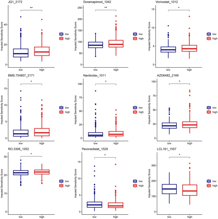
▲ Drug sensitivity profiles of MRS subgroups. The boxplots display the comparison of imputed sensitivity scores for nine candidate drugs (JQ1_2172, Doramapimod_1042, Vorinostat_1012, BMS_754807_2171, Navitoclax_1011, AZD6482_2169, RO_3306_1052, Pevonedistat_1529, and LCL161_1557) between the "low" and "high" MRS risk subgroups. The data indicate that the high-risk subgroup exhibits significantly higher sensitivity scores for LCL161_1557 compared to the low-risk subgroup. *P < 0.05; **P < 0.01.
LCL161 (T2080) was included among the candidate anti-tumor drugs evaluated in a lung adenocarcinoma study that stratified samples using a manganese metabolism–related risk score (MRS). Using this risk model, the authors performed drug susceptibility analyses to compare responses across subgroups. Within this framework, the high-MRS group was reported to be more responsive to LCL161 in the drug susceptibility analysis. Consistent with this finding, the study’s drug sensitivity analyses suggested that LCL161 may be preferable for individuals categorized into the high-MRS subgroup. These results position LCL161 within the study as a compound showing comparatively greater predicted sensitivity in the high-risk stratum defined by manganese metabolism–related gene expression. The evidence is based on computational drug susceptibility profiling tied to the constructed MRS and indicates a differential response pattern in which LCL161 aligns with the response profile of the high-MRS cohort. The analysis thereby highlights LCL161 as one of the compounds with greater expected activity in this specific molecularly defined risk group.[3]
■ 2.2 KIF5B-driven unfolded protein response reprograms breast cancer immunosuppressive microenvironment for single-cell guided therapeutic targeting.

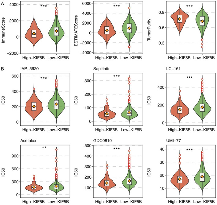
▲ Immune features and drug prediction of KIF5B. (A) Violin plots show the correlation between KIF5B expression groups (High-KIF5B vs. Low-KIF5B) and Immune Score, ESTIMATE Score, and tumor purity; the Low-KIF5B group exhibits significantly higher Immune Score and ESTIMATE Score, along with lower tumor purity. (B) Drug sensitivity prediction based on KIF5B groups. Among the six candidate drugs, the Low-KIF5B group exhibits significantly higher predicted IC50 values for LCL161 compared to the High-KIF5B group, indicating stronger resistance to LCL161 (***P < 0.001). Significant inter-group differences are also observed in the sensitivity profiles for the other drugs (IAP-5620, Sapitinib, Acetalax, GDC0810, UMI-77). ***P < 0.001, **P < 0.01.
LCL161 (T2080) was highlighted by the study’s drug sensitivity analyses as a candidate with activity in a specific molecularly defined subgroup. Using oncoPredict, the authors conducted drug sensitivity prediction and identified LCL161 among compounds with potential efficacy in patients characterized by high KIF5B expression. The analysis further emphasized LCL161 as one of the agents that may be particularly effective in KIF5B-high patients, and the authors noted this finding as providing a rationale for future preclinical and clinical evaluation. Together, these computational predictions position LCL161 as a prioritized compound within the context of KIF5B-based stratification, supporting its consideration where elevated KIF5B expression defines the target population. The reported findings consistently point to LCL161’s predicted activity in this setting and frame it as a candidate for follow-up evaluation based on stratified drug sensitivity assessment.[4]
■ 2.3 BET inhibitor-based combinations targeting novel dependencies in MECOM-rearranged (r) AML.
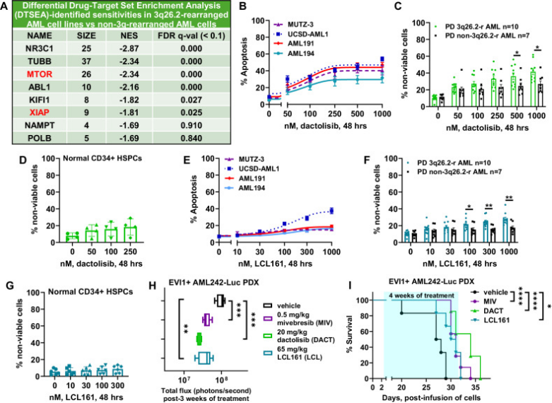
▲ (A) Table of target level, MECOM-r AML specific sensitivities identified from the drug screen shown by DTSEA-determined normalized enrichment score with corresponding FDR q-value < 0.1. (B) MUTZ-3, UCSD-AML1, AML191, and AML194 cells were treated with the indicated concentrations of dactolisib for 48 h. Cells were stained with annexin V-FITC and TO-PRO-3 iodide, and % apoptotic cells were analyzed by flow cytometry. Lines indicate the mean of three experiments; bars indicate standard error of the mean (S.E.M.). (C, D) Primary, patient-derived MECOM-r and non-MECOM-r AML or normal CD34+ HSPC samples were treated with the indicated concentrations of dactolisib for 48 h, followed by TO-PRO-3 iodide staining and flow cytometry analysis. * = p < 0.05 compared to non-MECOM-r AML cells. (E) MUTZ-3, UCSD-AML1, AML191, and AML194 cells were treated with indicated concentrations of LCL161 for 48 h, stained with annexin V-FITC and TO-PRO-3, and % apoptotic cells were analyzed by flow cytometry. (F, G) Primary, patient-derived MECOM-r and non-MECOM-r AML or normal CD34+ HSPC samples were treated with indicated concentrations of LCL161 for 48 h, followed by TO-PRO-3 staining and flow cytometry analysis. * = p < 0.05; ** = p < 0.01. (H) Total bioluminescence flux (photons/second) in NSG mice engrafted with luciferase-expressing MECOM-r AML242 xenografts and treated with mivebresib (0.5 mg/kg), dactolisib (20 mg/kg), or LCL161 (65 mg/kg) for 3 weeks. ** = p < 0.01; *** = p < 0.005 as determined by a two-tailed, unpaired t-test in GraphPad V10 software. (I) Kaplan-Meier survival plot of NSG mice engrafted with luciferase-expressing MECOM-r AML242 xenografts and treated for 4 weeks as indicated. * = p < 0.05; **** = p < 0.001 as determined by a log rank sum test in GraphPad V10 software.
LCL161 (T2080) is reported as an IAP antagonist. In cellular assays, LCL161 induced apoptosis in MUTZ-3, AML191, and AML194 cells. UCSD-AML1 cells were relatively more sensitive to the lethal effects of LCL161. Mechanistically, Western analysis showed that LCL161 reduced c-IAP1 protein levels in AML191 and UCSD-AML1 cells. Consistent with enhanced apoptotic signaling, greater sensitivity of UCSD-AML1 cells to LCL161-induced apoptosis was associated with higher induction of cleaved caspase-3, caspase-7, and PARP compared to dactolisib in UCSD-AML1 versus AML191 cells. In primary, patient-derived samples, LCL161 induced greater lethality in AML cells harboring MECOM rearrangements relative to those without these rearrangements, while LCL161 treatment spared normal CD34+ hematopoietic stem/progenitor cells. In an in vivo MECOM-rearranged AML242 xenograft model in NSG mice, monotherapy with LCL161 significantly reduced AML burden and improved survival compared to vehicle controls. In combination studies across four MECOM-rearranged AML cell lines, co-treatment with mivebresib and LCL161 induced synergistic lethality with delta synergy scores greater than 1.0 in all tested lines.[5]
■ 2.4 SMAC mimetics induce human macrophages to phagocytose live cancer cells.

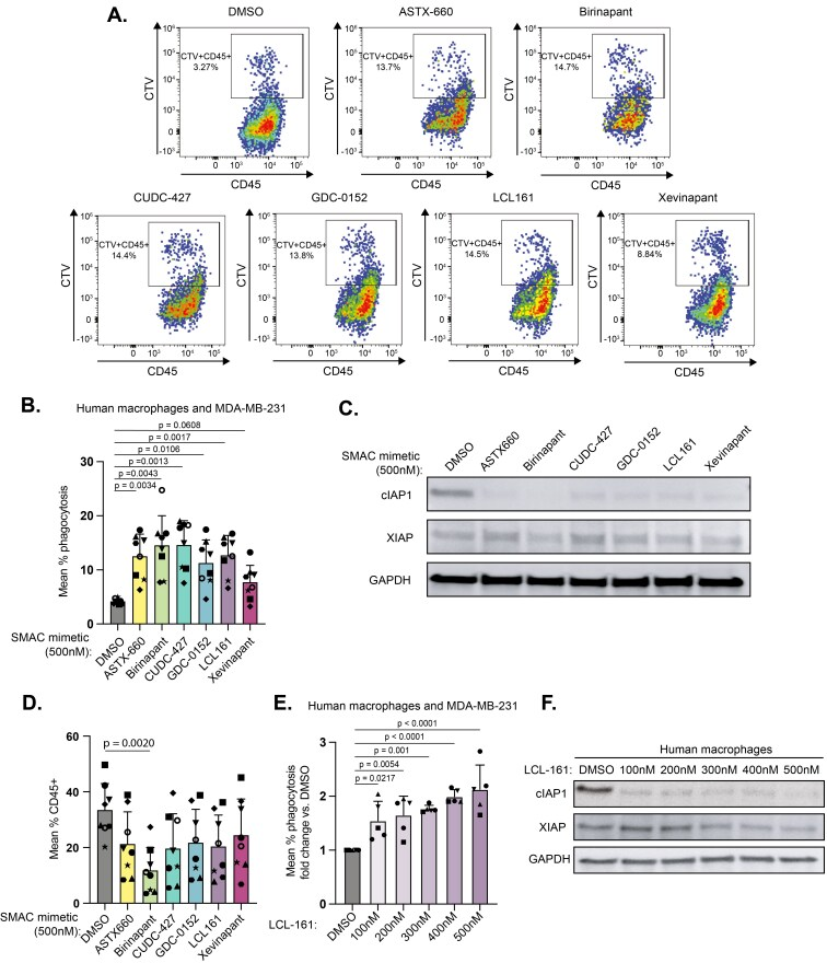
▲ (A) Representative flow cytometry plots from a single donor showing the CTV⁺CD45⁺ phagocytosis population within the total CD45⁺ macrophage population. (B) Macrophages from n=8 healthy donors were treated with 500 nM of the indicated SMAC mimetics for 24 hours prior to co-culture with CellTrace Violet-labeled MDA-MB-231 breast cancer cells. Phagocytosis was assessed by flow cytometry after 18 hours of co-culture. Results from each donor were normalized to the vehicle control from that donor. Error bars are SD. Statistical significance was determined using a pairwise one-way ANOVA with Sidak’s multiple comparison. (C) Immunoblot analysis of macrophage lysates treated with SMAC-mimetics (500 nM) for 24 hours. Blots were probed for cIAP1, XIAP, and GAPDH. (D) The frequency of macrophages recovered after 18 hours of co-culture with tumor cells under each of the indicated treatment conditions. (E) Macrophages from n=5 healthy donors were treated with the indicated concentrations of LCL161 for 24 hours prior to co-culture with CellTrace Violet-labeled MDA-MB-231 breast cancer cells. Phagocytosis was assessed by flow cytometry after 18 hours of co-culture. Results from each donor were normalized to the vehicle control from that donor. Error bars are SD. Statistical significance was determined using an ordinary one-way ANOVA. (F) Immunoblot of protein lysates from macrophages treated for 24 hours with the indicated concentrations of LCL161.
LCL161 (T2080) led to autocrine production of TNFa by cancer cells and T cells. In macrophages, LCL161 induced phagocytosis, as evidenced by the observation that genetic loss of TNFa caused complete ablation of LCL161-induced phagocytosis, indicating a TNFa-dependent phagocytic phenotype. Transcriptionally, LCL161 produced a detectable shift in macrophage state, with an LCL161-induced increase along a principal component analysis axis. Differential expression analysis showed that LCL161 alone induced a defined subset of NF-kB pathway genes, including MAP3K14, TRAF1, NFKB2, and NFKBIE. When combined with IFNg, LCL161 was associated with a robust interferon-responsive gene program, exemplified by increased expression of JAK2, CXCL9, and CXCL10. Together, these findings indicate that LCL161 promotes a TNFa-dependent phagocytic phenotype in macrophages and drives specific transcriptional changes consistent with NF-kB activation alone and a strong interferon response when used with IFNg.[6]
■ 3. Conclusion
LCL161 functions as a SMAC mimetic that antagonizes inhibitor of apoptosis proteins (IAPs) by mimicking the AVPI motif of endogenous SMAC/DIABLO, binding to BIR domains of XIAP and cIAP1. This releases caspases from inhibition and shifts TNFR1 signaling from pro-survival to pro-apoptotic complexes, promoting caspase activation and cell death. Literature findings demonstrate LCL161’s potent inhibition of XIAP and cIAP1 with nanomolar IC50 values, its ability to sensitize various cancer cell lines (including AML with MECOM rearrangement) to apoptosis, and its role in inducing TNFα-dependent macrophage phagocytosis. Computational drug sensitivity analyses further indicate LCL161’s selective efficacy in molecularly defined high-risk subgroups such as high manganese metabolism-related risk score (MRS) and high KIF5B expression cohorts. Future research may explore LCL161’s therapeutic potential in stratified patient populations, combination regimens with agents like BET inhibitors, and its immunomodulatory effects to enhance anti-tumor immunity.
■ 4. Q&A
Q1: What is the primary mechanism by which LCL161 induces apoptosis?
A1: LCL161 mimics the AVPI motif of SMAC/DIABLO to bind IAP BIR domains, releasing caspases from XIAP inhibition and promoting cIAP1 auto-ubiquitination and degradation, thereby shifting TNFR1 signaling towards apoptotic pathways and enhancing caspase activation.
Q2: In which molecularly defined patient subgroups has LCL161 shown predicted higher efficacy?
A2: LCL161 has shown predicted higher efficacy in patients with high manganese metabolism-related risk scores (MRS) and in those with high KIF5B expression, according to computational drug sensitivity analyses.
Q3: How does LCL161 affect macrophages according to the studies?
A3: LCL161 induces a TNFα-dependent phagocytic phenotype in human macrophages, activating NF-κB pathway genes and, when combined with IFNγ, triggering a strong interferon-responsive gene expression profile.
■ 5. Reference
[1] Fulda S, Vucic D, et al. Targeting IAP proteins for therapeutic intervention in cancer. Nat Rev Drug Discov. 2012;11(2):109-124.
[2] Vince JE, Wong WW-L, et al. IAP antagonists target cIAP1 to induce TNFα-dependent apoptosis. Cell. 2007;131(4):682-693.
[3] Li H, Wei S, Li W, Sun J, Wang Y. Integration of single-cell and bulk transcriptomics reveals the association of manganese metabolism-related genes with prognosis and immune infiltration in lung adenocarcinoma.. Clinical & translational immunology. 2026;15(3):e70085.
[4] Liu Y, Li S, Luo Z, Xia Y, Zhang P. KIF5B-driven unfolded protein response reprograms breast cancer immunosuppressive microenvironment for single-cell guided therapeutic targeting.. Discover oncology. 2026;17(1):333.
[5] Birdwell C, Fiskus W, Mill C, Kadia T, Daver N, DiNardo C, et al.. BET inhibitor-based combinations targeting novel dependencies in MECOM-rearranged (r) AML.. Leukemia. 2026;40(2):304-313.
[6] Liu S, Hulsman M, Leyendecker P, Chang E, Donovan K, Strobel F, et al.. SMAC mimetics induce human macrophages to phagocytose live cancer cells.. Immunotherapy advances. 2025;5(1):ltaf026.
https://www.targetmol.com/
TargetMol Chemicals Inc.


